An Ultrasound is a safe and painless diagnostic imaging technique based on the application of ultrasound. It is used to create an image of internal body structures such as tendons, muscles, joints, blood vessels, and internal organs. Its aim is often to find a source of a disease or to exclude pathology. The practice of examining pregnant women using ultrasound is called obstetric ultrasound and was an early development and application of clinical ultrasonography.
Ultrasound refers to sound waves with frequencies which are higher than those audible to humans. Ultrasonic images, also known as sonograms, are made by sending pulses of ultrasound into tissue using a probe. The ultrasound pulses echo off tissues with different reflection properties and are recorded and displayed as an image.
Compared to other dominant methods of medical imaging, ultrasound has several advantages. It provides images in real-time and is portable and can be brought to the bedside.
Ultrasound examinations can help to diagnose a variety of conditions and to assess organ damage following illness.
Ultrasound is used to help physicians evaluate symptoms such as:
Ultrasound is a useful way of examining many of the body’s internal organs, including but not limited to the:
Ultrasound is also used to:
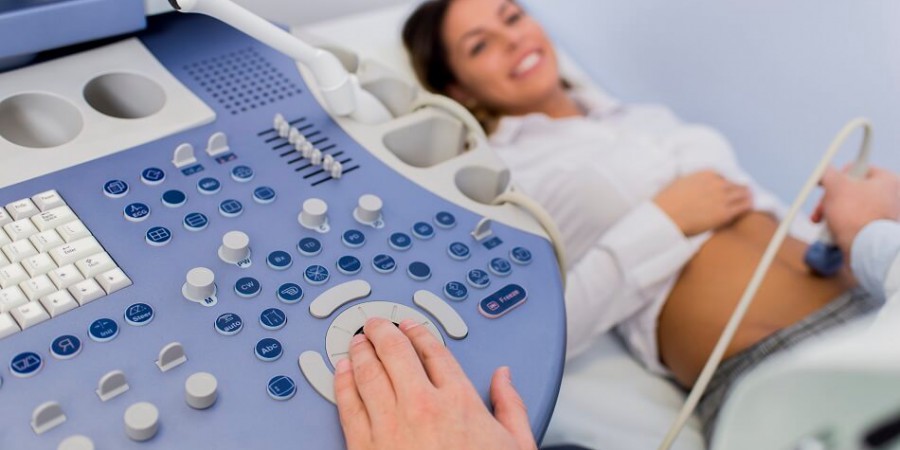
Obstetric ultrasound produces pictures of a baby within a pregnant woman, as well as the mother’s uterus and ovaries. It is used for monitoring pregnant women and their unborn babies. A Doppler ultrasound study is a technique used to evaluate the blood flow in the umbilical cord, fetus or placenta.
Ultrasound imaging of the abdomen produces pictures of the structures within the upper abdomen. It is used to help diagnose pain or distention and evaluate the kidneys, liver, gallbladder, bile ducts, pancreas, spleen and abdominal aorta. Ultrasound is safe, and noninvasive.
Ultrasound imaging of the pelvis produces pictures of the structures and organs in the lower abdomen and pelvis. There are three types of pelvic ultrasound: abdominal, vaginal (for women), and rectal (for men). These exams are frequently used to evaluate the reproductive and urinary systems.
Vascular ultrasound evaluates the body’s circulatory system and help identify blockages in the arteries and veins and detect blood clots. A Doppler ultrasound study evaluates blood flow through a blood vessel. Ultrasound provides images of soft tissues that don’t show up on x-ray images.
Ultrasound imaging of the breast produces pictures of the internal structures of the breast. It is primarily used to help diagnose breast lumps or other abnormalities your doctor may have found during a physical exam, mammogram or breast MRI. Ultrasound is safe, noninvasive and does not use radiation.
Thyroid ultrasound produces pictures of the thyroid gland within the neck. It is commonly used to evaluate lumps or nodules found during a routine physical or other imaging exam.

.png)


.svg)